| 产品名称 | BIOMIMESYS Liver,BIOMIMESYS Hydroscaffold,BIOMIMESYS?肝脏三维培养板,BIOMIMESYS?肝脏细胞外微环境 | ||||||||||||||||||||||
| 品牌 | biomimesys | ||||||||||||||||||||||
| 产品货号 | BIOMIMESYS Liver,BIOMIMESYS Hydroscaffold,BIOMIMESYS?肝脏三维培养板,BIOMIMESYS?肝脏细胞外微环境 | ||||||||||||||||||||||
| 产品价格 | 现货询价 | ||||||||||||||||||||||
| 联系人 | 李先生 | ||||||||||||||||||||||
| 联系电话 | 18618101725 | ||||||||||||||||||||||
| 产品说明
BIOMIMESYS® is an organ-specific Extracellular Matrix (ECM) formed by the crosslinking reaction of hydrosoluble modified hyaluronic acid (HA) and other ECM components (collagens, fibronectin, etc.) with ADH (Adipic acid dihydrazide), to create a "Hydroscaffold" 3D cell culture system. BIOMIMESYS® Liver represents a new generation of mimetic hydroscaffolding for 3D hepatocyte-like cell cultures. Available in a ready-to-use format, it enables the culture of hepatocytes and hepatocyte-like cells under physiological conditions that are representative of the microenvironment found in liver tissue. The highly porous nature of the hydroscaffold allows the rapid uptake of nutrients, oxygen, etc. into the cells to create a reproducible study model for all downstream analyses used with 3D hepatocytes-like cells culture (metabolism, toxicity, etc.).
BIOMIMESYS® Liver is easy & ready-to-use. Upon receiving the vacuum sealed 96-well plate open it (under a hood) and add the cells directly on top of the matrix. Changing the culture medium is easy as well. To remove medium, simply draw the medium with a pipette between the matrix and the edge of the well. To refresh the medium, place fresh medium onto the surface of the matrix.
Composition
Composed of hyaluronic acid (HA) - a major component of the cell’s extracellular matrix (ECM) - biofunctionalized with liver ECM components (Collagens type I and IV, galactosamine and cell binding domain of Fibronectin), the BIOMIMESYS®Liver hydroscaffold for 3D hepatocyte cultures is thus an ideal study model because it is;
? Representative of the hepatocytes' microenvironment in terms of cell-cell interactions, matrix composition and cell-matrix interactions
? Highly porous, enabling easy analysis of supernatant (for toxicity and metabolism studies)
? Transparent for direct visualization of cells
Composition of the Scaffold
? Hyaluronic acid (HA) - a major component of the cell’s extracellular matrix (ECM)
PHYSICOCHEMICAL FEATURES CELLS TESTED
BIOMIMESYS® Liver: Liver Extracellular Microenvironment
Like most cells, the hepatocyte microenvironment plays an important role in the maintenance of differentiation, polarization and cellular functions. The extracellular matrix (ECM) is a complex mixture of functional and structural proteins, glycoproteins and glycosaminoglycans (GAG) organized in a tissue-specific three-dimensional structure. Numerous studies have shown that using de-cellularized organs for hepatocyte culture is integral for recreating a microenvironment favorable to the survival and maintenance of cell functions (3, 4).
The liver extracellular microenvironment notably contains collagen I, collagen IV and fibronectin, all of which play a key role in the structuring of tissue and maintenance of hepatocyte functions (5).
To mimic this bioactive microenvironment, HCS Pharma has developed the world’s first biomimetic hydroscaffold of HA, collagen type I and IV, galactosamine and RGDS motif of fibronectin dedicated to the culture of hepatocytes: BIOMIMESYS® Liver.

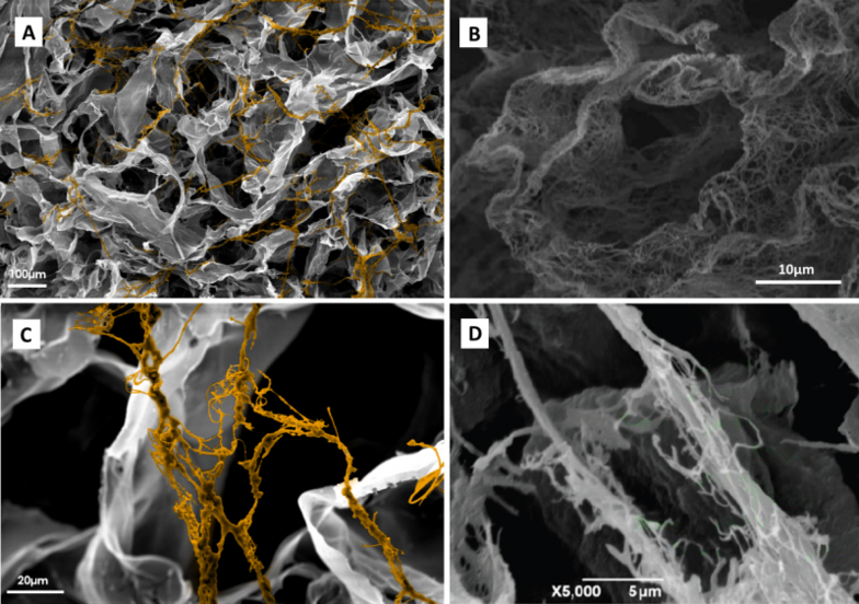
Fig. Scanning Electron Microscopy of BIOMIMESYS®Liver showing collagen fibers, artificially stained (A and C), decellularized human liver (B, ref 5) and collagen fiber in decellularized rat liver (D, ref 6).
Cell cultures in BIOMIMESYS®Liver were performed with the HepG2 cell line and cryopreserved human primary hepatocytes (KalyCell).
Seeding was performed following the protocol described in the “first steps document” with the following cell densities and volumes:
? Primary Hepatocytes: 25 μl with 75,000 cells / gel
? HepG2: 25 ?l with 50 000 cells / gel
Biomimesys and Hepatocyte Formation
The liver is composed of various cell types, 60% of parenchymal cells or hepatocytes and 40% non-parenchymal cells (1).
Hepatocytes are highly polarized cells with an apical domain representing about 10 to 15% of the cell surface. This domain is composed of a canalicular network formed between two adjacent hepatocytes connected by tight junctions. These bile ducts are approximately 1?m in diameter and carry bile acid secreted by hepatocytes.
The hepatocytes are highly complex cells providing a variety of functions, the main ones are grouped in the term “ADMET” (Absorption, Distribution, Metabolism, Excretion, Transport). Hepatocytes control homeostasis and the toxicity of molecules, as they absorb these molecules from blood to metabolize and redistribute them to various organs. Hepatocytes are also involved in the processing or degradation of molecules with therapeutic purposes that can generate potentially toxic secondary compounds.
During the development phase, about 40% of new drug candidates are discarded due to toxicity (2). Early detection of this toxicity not only saves money and time, but also decreases potential harm to animal test subjects.
Biomimesys guarantees the polarization of hepatocytes essential for biliary secretion and proper cellular function. It enables the culture of hepatocytes under physiological conditions that are representative of the microenvironment found in liver tissue. The advantages of Biomimesys Liver compared to traditional 2D culture can be seen below:
? Conventional 2D culture: Shows canalicular spheres between adjacent cells, but no canalicular network.
? Collagen sandwich culture (2D): A multicellular canalicular network can be seen. Viabile for up to 2 weeks.
? BIOMIMESYS® LIVER: Reported greater amount of Biliary Canaliculi formation, higher CYP activities and Albumin Secretion, and increased culture time than 2D culture.
| |||||||||||||||||||||||